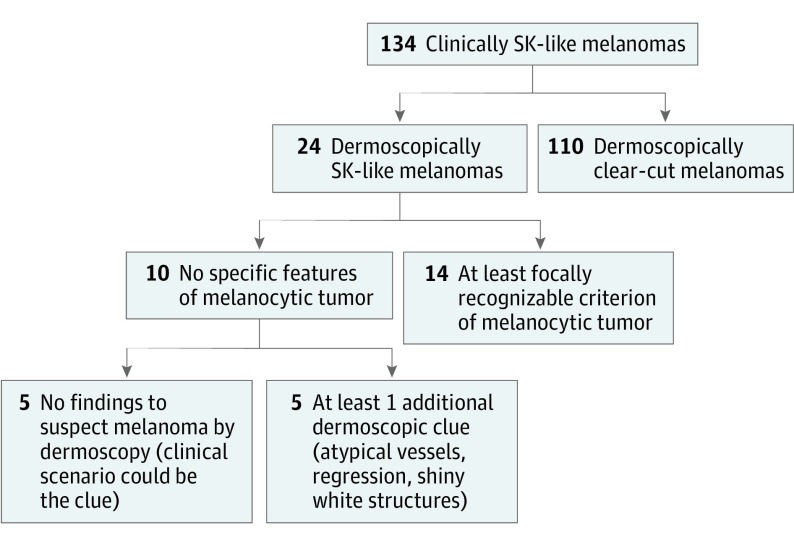
Figure 1. Diagram of Seborrheic Keratosis (SK)–like Melanoma Detection by Different Dermoscopic Clues.
A total of 134 clinically SK-like melanomas were evaluated; among them, 24 cases (17.9%) were considered possible SK (dermoscopically SK-like melanomas). Of these, 14 showed at least 1 recognizable criterion of a melanocytic lesion: pigment network, globules or dots, pseudopods, blue-white veil, and/or the blue-black sign.

