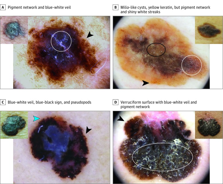
Figure 2. Four Seborrheic Keratosis (SK)–like Melanomas Belonging to the Group of 110 Lesions Easily Detected by Dermoscopy.
Insets, Pigmented lesions with some degree of hyperkeratotic surface and sharp demarcation that clinically can simulate SKs. Larger images, Dermoscopy shows features suggestive of melanocytic lesions and therefore melanoma. A, Presence of pigment network at the periphery (arrowhead) with hyperkeratosis and blue-white veil in the center (circle). B, Brownish lesion with notable milia-like cysts (black oval) and yellowish keratin, pigment network (arrowhead), irregular globules and dots, and shiny white streaks (white circle). C, Blue-white veil and the blue-black sign in the center, in addition to atypical network (black arrowhead) and pseudopods at the periphery (blue arrowhead). D, Markedly hyperkeratotic tumor with verruciform surface and blue-white veil (white oval); the clue is at the periphery, with atypical network (arrowhead) and regression.

