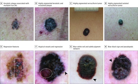
Figure 3. Four Seborrheic Keratosis–like Melanomas Needing Careful Dermoscopic Evaluation to Be Correctly Diagnosed.
A-D, Clinical photographs. In B and C, the rules are in millimeters. E-H, Dermoscopic images of the same lesions. Pigmented lesions with marked hyperkeratotic surface partially impeding the easy observation of the dermoscopic clues. The proper use of immersion liquid and enough light may help the evaluation (compare G, which did not have enough liquid, and H, which is a proper image that allows the detection of the pigment network [circle]). The presence of subtle pigment network (arrowheads in G and H) and globules (arrowhead in F) at the periphery of the lesions and the blue-black sign are the main key features. Regression features (box in E; circle in F) can be another clue to recommend excision.

