
Figure 1.
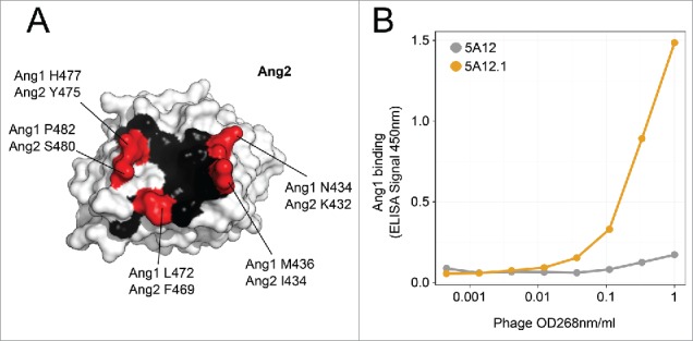
(A) Structural epitope of 5A12 on Ang2 as observed in 5A12:Ang2 crystal structure (PDB 4ZFG) is mapped onto the surface of Ang2. Epitope positions (< 5Å from 5A12) that are conserved between Ang1 and Ang2 are colored in black, while positions that differ between the two proteins are labeled red. (B) Phage titration ELISA to measure the binding of phage displaying either the parental Ang2/VEGF DAF 5A12 (gray) or the affinity maturated version 5A12.1 (orange) to Ang1.