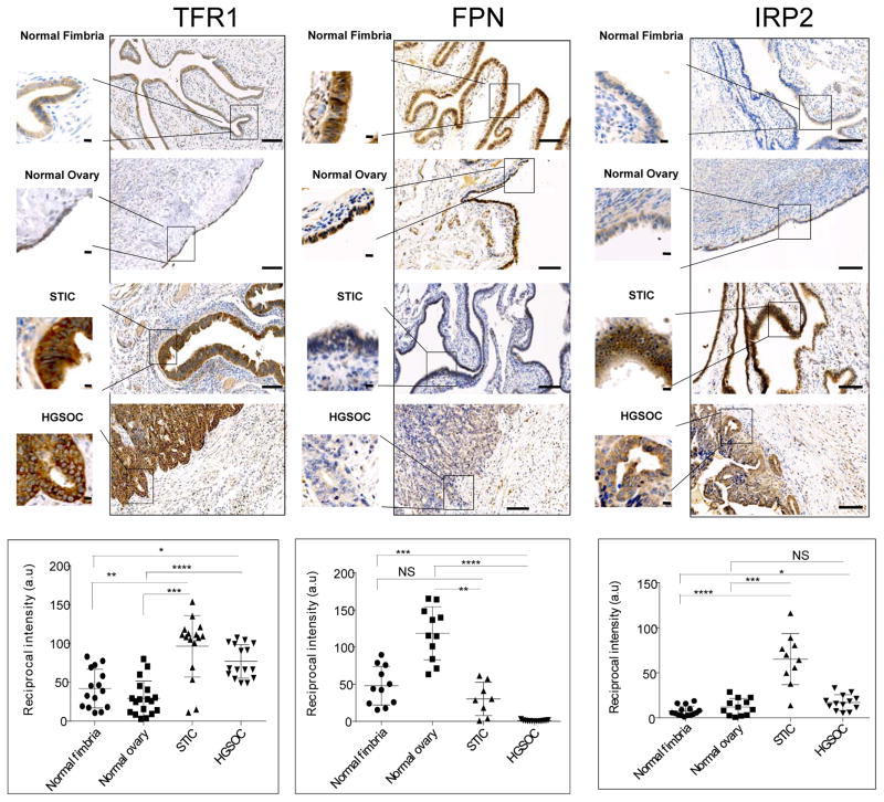
Fig. 1. Proteins that control intracellular iron are altered high grade serous ovarian cancer (HGSOC).
Representative images of immunohistochemical staining of normal fimbria, normal ovary surface epithelia, serous tubal intra-epithelial carcinoma (STIC) and HGSOC stained with antibodies against transferrin receptor 1 (TFR1), ferroportin (FPN) and iron regulatory protein 2 (IRP2). Dot plots represent quantification of staining of tissues collected from 8 patients with HGSOC and 5 with STIC compared to 8 subjects with normal fimbria and 6 individuals with normal ovarian surface epithelium. Images of three to four random fields per slide were quantified. Differences in TRFC (p<0.0001), FPN (p<0.0001 and IRP2 (p<0.01) were statistically significant (one way ANOVA). *p<0.001, **p<5x10⁻⁵, *** p<5x10⁻⁶, ****p<5x10⁻⁷, one tailed t test for individual comparisons. Scale bar 1 mm; inset scale bar 10 μm.

