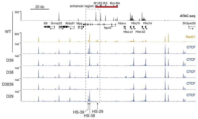
Figure 3. Disruption of CTCF binding motifs results in loss of CTCF binding at the α-globin locus in primary erythroid cells derived from mutant mice.
ATAC-Seq (RPKM) in wild-type (WT) primary erythroid cells shows chromatin accessibility across the α-globin locus. Local genes (Refseq) and the α-globin enhancers are annotated. Normalised CTCF ChIP-seq reads (RPKM, 2 independent experiments in which 2 animals were analysed in total) across the α-globin locus are shown for WT and each of the generated CTCF binding site mutants; D39: HS-39 mutant, D38: HS-38 mutant, D3839: combined HS-38 and HS-39 mutant, D29: HS-29 mutant. Also shown is normalised Rad21 ChIP-seq (RPKM, 2 independent experiments in which 2 animals were analysed in total) for WT primary erythroid cells. The dashed box indicates the genomic region within which the generated CTCF binding mutations are located.

