
FIGURE 11.
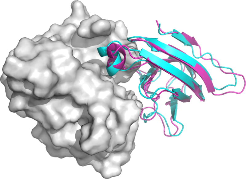
Docking the E. coli PliG lysozyme inhibitor to the salmon goose-type lysozyme using SAXS data as restraints. A near-native model (shown cyan cartoon) was obtained as the center of the 3rd largest cluster. The X-ray conformation of the inhibitor is shown as magenta cartoon.