
FIGURE 13.
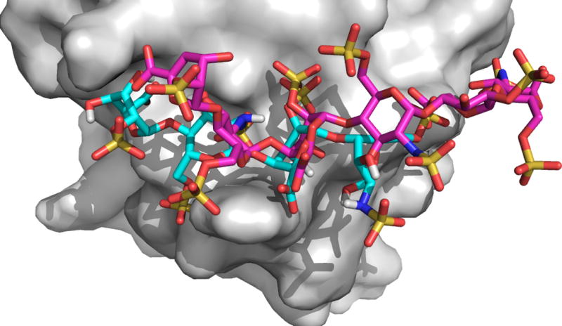
Docking of the heparin tetramer probe to the ligand-free structure of the basic fibroblast growth factor. The center of the second largest cluster is shown as cyan sticks. The X-ray structure of the bound hexamer shown in magenta.
Official websites use .gov
A
.gov website belongs to an official
government organization in the United States.
Secure .gov websites use HTTPS
A lock (
) or https:// means you've safely
connected to the .gov website. Share sensitive
information only on official, secure websites.
Docking of the heparin tetramer probe to the ligand-free structure of the basic fibroblast growth factor. The center of the second largest cluster is shown as cyan sticks. The X-ray structure of the bound hexamer shown in magenta.