
FIGURE 8.
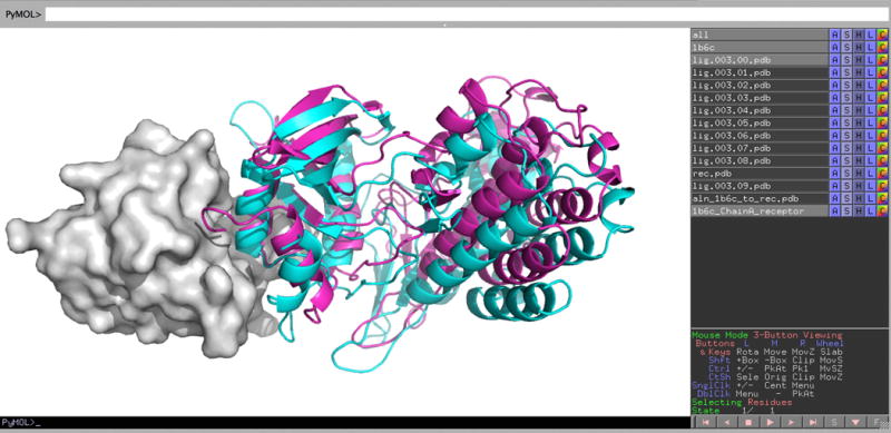
Screen image of exploring the results of running ClusPro in Others Mode. Results are shown using PyMOL from docking the X-ray structure of the ligand, to the X-ray structure of the FK506 binding protein (FKBP). The receptor, FKBP, is shown as grey surface, and the ligand (lig.003.00.pdb) at center of the largest cluster is shown as cyan cartoon. For comparison we superimposed the native complex (PDB ID 1B6C) on the receptor in the docked structure, and the corresponding ligand pose is shown as magenta cartoon.