
FIGURE 9.
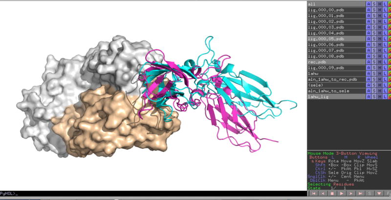
Screen image of the PyMOL visualization of the results of running ClusPro in Antibody Mode. We docked the X-ray structure of the extracellular domain of the human tissue factor (PDB ID 1TFH) to the unbound X-ray structure of the FAB domain of the inhibitory antibody 5G9 (PDB ID 1FGN). Both the heavy and light chains were used to represent the receptor. The center of the 6th most populated cluster, lig.000.05.pdb, is shown as cyan cartoon, whereas the antibody is shown in surface representation. The antigen in the native complex (PDB ID 1AHW) is shown as magenta cartoon. The IRMSD between native and predicted ligand poses is 4.7 Å.