
Fig. 1. BmP01 directly activates TRPV1 channel.
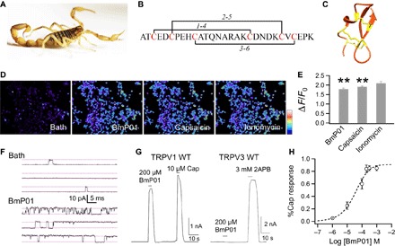
(A) Image of the scorpion M. martensii. (B) Peptide sequence and disulfide bonds of BmP01. (C) Solution structure of BmP01 [Protein Data Bank (PDB) ID: 1WM7]. Three disulfide bonds are colored in yellow. (D) Calcium imaging of hTRPV1-expressing HEK293 cells challenged sequentially with BmP01 (200 μM), capsaicin (10 μM), and ionomycin (1 mM). The scale bar represents 126 to 314 arbitrary units. (E) Fluorescence signals of hTRPV1-expressing HEK293T cells in response to BmP01, capsaicin, and ionomycin. **P < 0.001 (n = 3 to 5). (F) Representative single-channel current traces recorded in the absence or presence of 100 μM BmP01 (pH 7.0). (G) Representative whole-cell current response to BmP01 and agonists at saturating concentration [for hTRPV1, 10 μM capsaicin; for mTRPV3, 3 mM 2-aminoethoxydiphenyl borate (2APB)]. (H) Concentration-response relationship of BmP01 and hTRPV1 (n = 10) fitted to a Hill equation with the following parameters: EC50, 40.4 ± 12.3 μM; slope factor, 0.80 ± 0.05.