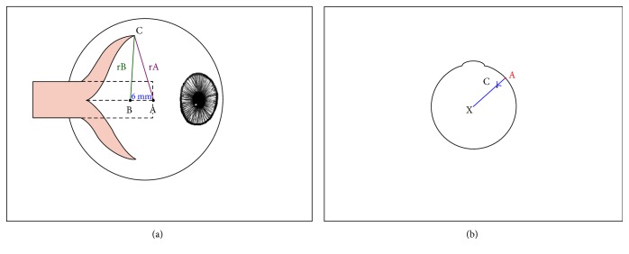
Figure 1.
Schematic representation of the Y-splitting procedure of the medial rectus muscle. (a) Side view. (A) point at the middle of the original insertion (dotted line) of the medial rectus muscle. (B) point 6 mm distal of point A. (rA, rB) radius of predetermined distances (see text). Calipers are centered on point A, and a circle is drawn on the sclera with rA; then, the calipers are centered on point B, and a circle with rB is marked on the sclera. The intersection of the circles marks point C, the point of scleral refixation. In this graph, measurements are shown for reattachment of the superior muscle section; the same is applied to the lower half. (b) View of the eye from above. Reattaching the split medial rectus to point C (shown for the superior half) reduces the lever arm (the distance between the center of the globe and the insertion of the muscle), and thus the torque.

