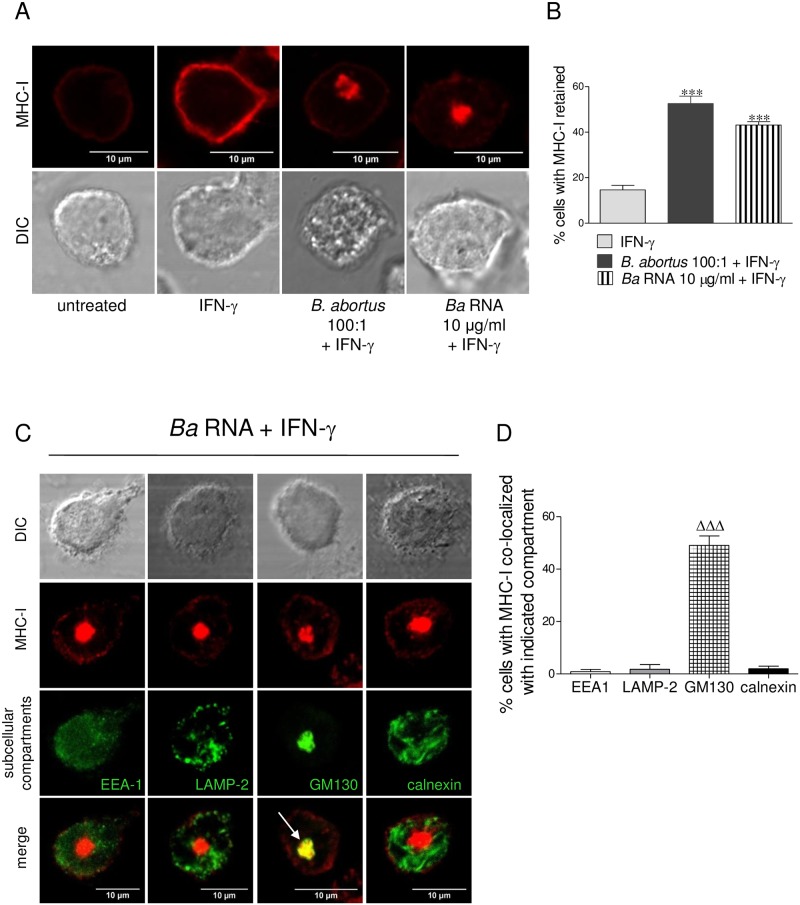
Fig 7. B. abortus RNA mimics MHC-I intracellular retention in Golgi apparatus mediated by B. abortus infection.
(A) Confocal micrographs of THP-1 cells infected with B. abortus or treated with B. abortus RNA in the presence of IFN-γ for 48 h. MHC-I expression was determined with a primary anti-human MHC-I Ab (W6/32) and Alexa 546-labelled secondary Ab (red). (B) Quantification of MHC-I retention. Data are expressed as percentage of cells with MHC-I retained ± SEM of three independent experiments. The number of cells counted per experimental group was 200. (C) Confocal micrographs of THP-1 cells treated with B. abortus RNA in the presence of IFN-γ for 48 h. MHC-I expression was determined with a primary anti-human MHC-I Ab (W6/32) and Alexa 546-labelled secondary Ab (red). Subcellular localization markers were detected using mAbs specific for EEA1 (early endosomes), LAMP-2 (late endosomes/lysosomes), GM130 (Golgi) and calnexin (ER) followed by Alexa 488-labelled secondary Ab (green). White arrow shows co-localization (yellow staining). Results are representative of three independent experiments. (D) Quantification of co-localization of MHC-I with the subcellular compartments. Data are expressed as percentage of cells with MHC-I co-localized with indicated compartment ± SEM of three independent experiments. The number of cells counted per experimental group was 200. ***P<0.001 vs. IFN-γ-treated; ΔΔΔP<0.001 vs. the other subcellular compartments.

