
Fig. 4.
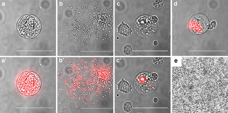
Representative microscopic images of detached cells. a Bright field and a′ fluorescence merged image of a normal detached cell, b and b′ of a ruptured detached cell and c, d examples of detached cells with abnormal morphology, c, c′ of merofusosomes [25] and d of absent merozoite formation. e Example of a fungal contamination of Plasmodium infected cells. Scale bars are 50 µm