Fig. 3.

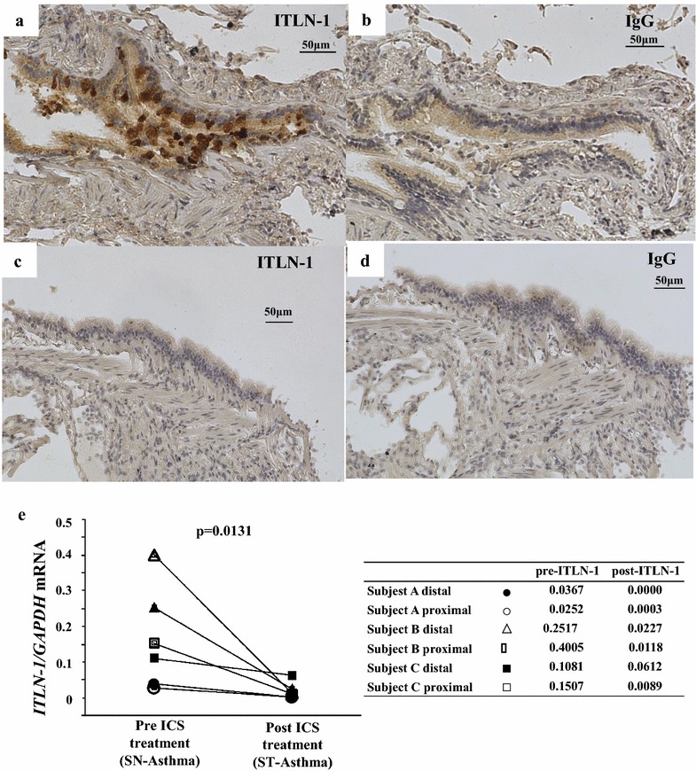
Immunohistochemistry of ITLN-1 expression in airway tissue. Representative distal BECs of SN-Asthma (a, b), and ST-Asthma (c, d) samples were stained with anti-ITLN-1 antibody (a, c) and IgG isotype control (b, d). The fields are 200 magnificent and antibodies are 1:500 diluted, respectively. ITLN-1 staining is mainly in the goblet cells in SN-Asthma. e ITLN-1 mRNA expressions before and after ICS (MF) treatment from 3 patients with 6 samples. Closed markers represent distal BECs and open markers are proximal BECs. Circles, triangles and squares are represented subjects individually. Additional small tables shows ITLN-1 mRNA values pre and post ICS treatments. Comparisons were done using the Wilcoxon test
