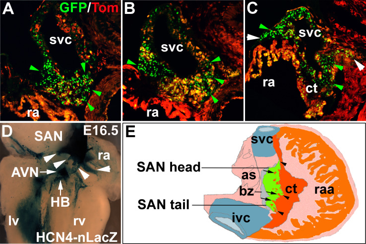
Fig. 1.
SAN structure. a–c Sections across SAN head, center and tail of Hcn4-nGFP, Nkx2–5-Cre and tdTomato triple positive mice showing pacemaker cell clusters of variable sizes (HCN4-GFP+, green arrowhead) intermingled with atrial strands (ast) (tomato+, Nkx2–5-Cre+) and potential sinus-atrial conduction paths (SACPs) (d, white arrowhead). d Wholemount Xgal staining of HCN4-nLacZ heart (endocardial view) showing cardiac conduction system, including SAN with potential SACPs (arrowhead). e A schematic diagram of SAN and the right atria (dorsal view), highlighting the interdigitation of SAN and surrounding atria working myocardium (ct), atria strands (arrow), potential SACPs (arrowhead) (as atrial septum, AVN atrioventricular node, bz block zone, ct crista terminalis, HB his-bundle, ivc inferior vena cava, lv left ventricle, ra right atria, raa right atrial appendage, rv right ventricle, SAN sinoatrial node, svc superior vena cava)

