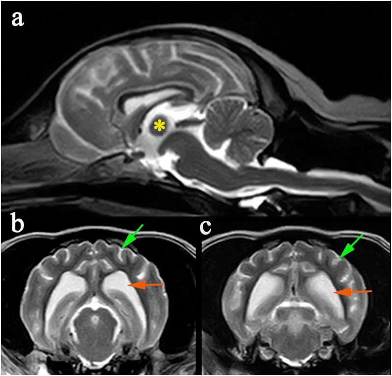
Fig 1. MRI changes in an 8 year old male neutered MWHD with advanced LD.
(a) Mid-sagittal T2W image of the brain demonstrating atrophy of the intra-thalamic adhesion (*). (b) Transverse T2W at the level of temporal lobes demonstrating cortical atrophy with widening of the subarachnoid space (green arrow) and enlargement of the lateral ventricle (orange arrow). (c) Transverse T2W at level of occipital lobes demonstrating cortical atrophy with widening of the subarachnoid space (green arrow) and enlargement of the lateral ventricle (orange arrow).

