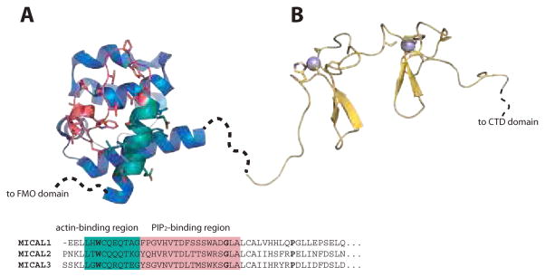
Figure 5. Structures of CH and LIM domains.
A. Solution structure of human MICAL1 CH domain (PDB 2DK9152) is shown as ribbons, highlighting the actin-binding region (cyan) and PIP2-binding region (pink) with sticks. The sequences of the N-terminal region of CH domains from mouse MICAL1–3 (see Figure 3) are detailed, mapping the conserved regions shown in the structure and the hydrophobic signature residues (bold). B. Solution structure of human MICAL1 LIM domain (PDB 2CO8) is shown as yellow ribbons, with cysteines and histidine involved in zinc atoms ligation (cyan spheres) represented as sticks. Figures is prepared with PyMOL, based on152,146.

