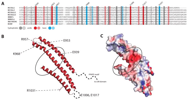
Figure 6. Conservation and structure of DUF3585 domains.
A. Alignment of the DUF3585 domains of mouse MICAL1 (NP-612188.1), MICAL3 (NP001257404.1), MICAL-L1 (NP803412.1), MICAL-L2 (NP777275.2), EHBP1 (NP001239444.1), EHBPL1 (NP001108069.1), EBITEIN1 (NP081863.2) and MINP (NP653101.1). Conserved residues are highlighted with red (acidic), blue (basic) or grey (hydrophobic) backgrounds. Full conservation is indicated with plain colors, while translucent colors indicated physicochemical conservation. B. The ribbon structure of human MICAL1 DUF3585 domain (PBD 5LPN103) is shown on the left side and its electrostatic surface on the right. Conserved residues (numbered accordingly to mouse MICAL1) are highlighted as stick, also indicated on part A (upper line). The conserved acidic patch is encircled. Figure was prepared with PyMOL.

