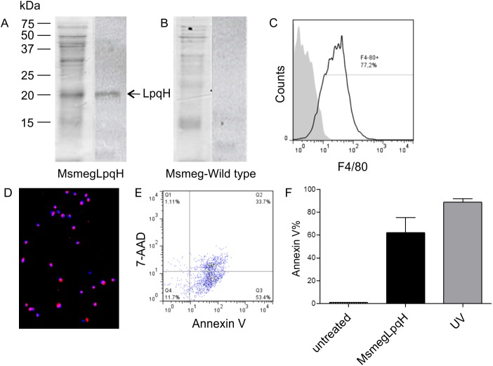
Fig 1. Mycobacterial cell wall proteins mediate apoptosis of bone marrow MØ.
A M. smegmatis strain was transformed to overexpress LpqH, a 19 kDA Mtb cell wall lipoprotein that is apoptogenic for MØs. The cell wall proteins of the transformed strain were disrupted with sonication and then separated by 15% SDS-PAGE. (A) A Coomassie blue stained gel is shown and LpqH (19 kDa) was detected by immunoblot with a mAb and HRP labeled rabbit antiserum. (B) Immunoblot failed to reveal LpqH in wild-type M.smegmatis. (C) Flow cytometry revealed that the great majority of MØs used in these assays were F4/80 positive. (D, E) Following the incubation of bone marrow-derived MØs with 50 μg cell wall protein for 24 h, high levels of apoptosis were revealed by immunofluorescence microscopy of TUNEL assays (excitation 496 nm, emission 575 nm) (original magnification, 20x) and by flow cytometry with Annexin V. A representative Annexin/ 7-AAD dot plot showed that 87.1% of MØs were apoptotic and 33.7% were necrotic. (F) Treatment of MØs with UV light also induced high levels of apoptosis.

