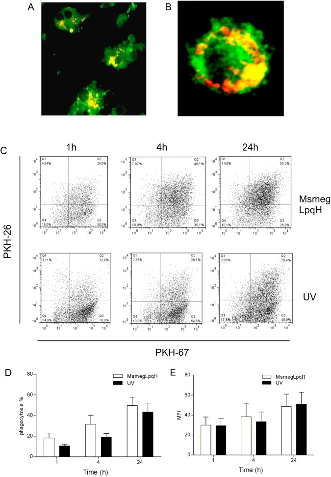
Fig 3. Inmature DCs efficiently phagocytose apoptotic MØ.
DC precursors were obtained from mouse bone marrow and were cultured in the presence of GM-CSF for 6 days. Phagocytosis assays were then conducted with PKH-67-labeled DCs and apoptotic PKH-26-labeled MØs. (A) With confocal microscopy, classical DC morphology was observed (original magnification, 60x). (B) In addition, fluorescencent apoptotic bodies appeared to reside within vacuolar structures, (original magnification, 100x) (PKH-67 excitation 493 nm, emission 525 nm; PKH-26 excitation 496, emission 575 nm). (C, D) Flow cytometry of the DC/MØ cocultures at various time points showed that phagocytosis increased with time and high levels of phagocytosis were detected 24 h after coculturing. (C, D, E) Phagocytosis of UV light induced apoptotic bodies was similar. The results shown are representative of three independent experiments. MFI, Mean fluorescence index.

