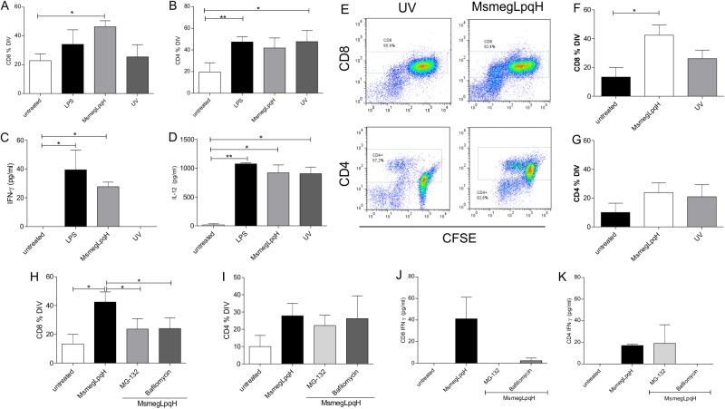
Fig 6. Induction of T cell proliferation by DCs activated with MsmegLpqH-induced apoptotic MØs.
To examine the capacity of the DCs that engulfed the apoptotic MØs to activate T cells, DCs were cocultured with autologous T cells isolated from the spleens of naïve mice by cell sorting with an anti-Thy mAb. DCs and isolated T cells were cocultured at a 1:10 ratio for 3 d. T cell proliferation was assessed by the CFSE dilution method. (A, B) DCs that phagocytosed the mycobacteria-induced apoptotic MØs triggered the proliferation of CD8+ T cells, and not CD4+ T cells (p ≤ 0.04, paired t Student’s test). (C, D) In the supernatants increased amounts of IFN-γ (p ≤ 0.01; Kruskall-Wallis test) and IL-12 (p ≤ 0.01 Mann Whitney test). (B, C, D) In comparison, the DCs activated with UV-induced apoptotic MØs promoted the proliferation of CD4+ T cells and the release of IL-12 (p ≤ 0.001, Mann Whitney test) and IFN-γ was within basal values. (E, F, G) Proliferation assays with CD4+ and CD8+ T cells isolated by cell sorting confirmed the ability of CDs activated with MsmegLpqH apoptotic MØs to trigger a proliferative response of CD8+ T cells (p ≤ 0.05, paired t Student’s test) but not of CD4+ T cells. To identify the pathways used by DCs to activate CD8+ T cells, the DCs were treated with the proteasome inhibitor, MG-132, and a vacuolar H+ ATPase inhibitor, bafilomycin. (H) Both inhibitors reduced the proliferation of the CD8+ T cells (p ≤ 0.01 and p ≤ 0.02, respectively, Dunn’s Multiple Comparison test). (I) In comparison the proliferation of CD4+ T cells was not inhibited. The results of four independent experiments are shown. (J, K) Also, a representative experiment shows that both inhibitors decreased the production of INF-γ by the CD8+ T cells but not by CD4+ T cells.

