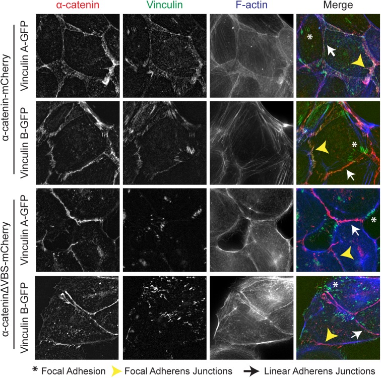
Fig 2. Zebrafish vinculin A and vinculin B localization.
Fixed α-catenin-depleted MDCK epithelial cells expressing either α-catenin-mCherry (top two rows, depicted in red) or α-cateninΔVBS-mCherry, which lacks the vinculin binding domain (bottom two rows, depicted in red). In addition, cells express zebrafish vinculinA-GFP or vinculinB-GFP (both depicted in green) and were stained for F-actin (blue). Asterisks mark Focal Adhesions, White arrows mark Focal Adherens Junctions and Yellow Arrowheads mark Linear Adherens Junctions.

