Fig. 8.

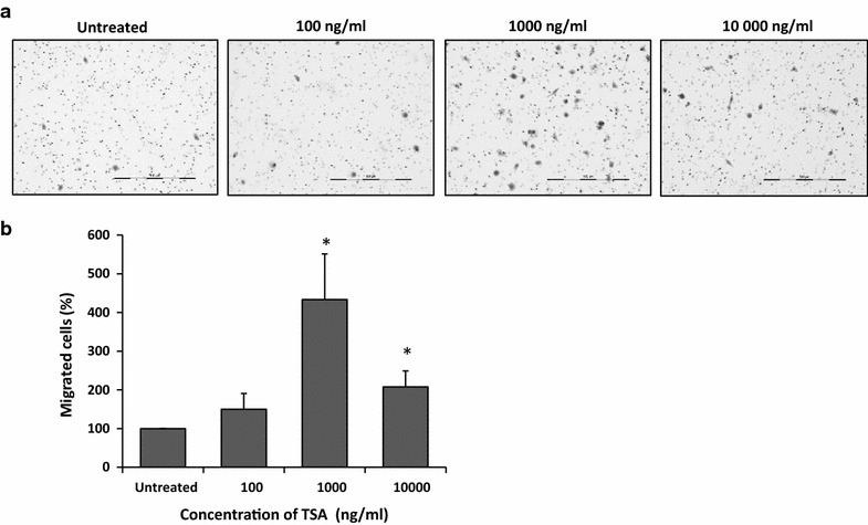
TSA enhanced migration of MCF-7 cells. Migration was assessed according to transwell migration assay. Migrated cells were viewed under inverted microscope (×10 magnification) and images were captured using a monochrome ProgRes CFcool CCD camera (Jenoptik, Germany). Migrated cells in each insert were counted and averaged from 30 random fields. a Representative images of migrated MCF-7 cells from one field view. b The percentage of migrated cells normalised to untreated. Data were collected from n = 3 independent experiments, presented as mean ± SEM. Unpaired Student’s t test *p < 0.05
