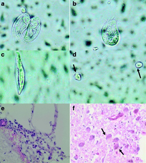
Fig. 1.

Different stages of N. fowleri in cerebral spinal fluid of a 24 year old Zambian male Different forms of Amoeba (a, b, c) and and cysts (d) in CSF. Fibrino-purulent material on the leptomeninges, consisting of lymphocytes, neutrophils and congested blood vessels (e) and Multiple N. fowleri amoeba (f) present in the brain parenchyma with hemosiderin pigment
