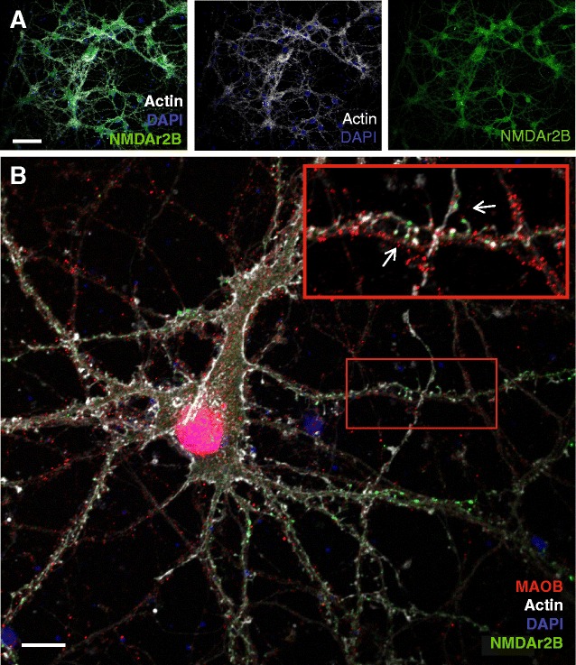
Fig. 3.

Immunocytochemistry in primary neurons to elucidate the MAO-B location in relation to the synapse. Primary hippocampal neurons were stained with Phalloidin to visualize fibrillar actin and the neuronal structure (white), DAPI to visualize nuclei (blue), anti-NMDAr2B to visualize the postsynaptic side of glutamatergic synapses (green) and anti-MAO-B (red). a Low-magnification overview imaged with a 20× objective, to visualize that most or all of the pyramidal neurons in our cell cultures contain NMDAr2B. b High-magnification image taken with a 60× oil immersion objective. Inset shows a zoomed part of the large image that was enriched with a high density of NMDAr-containing dendritic spines and MAO-B staining on the opposing axons. The intensity of the red channel in the inset was enhanced using image J (Color figure online)
