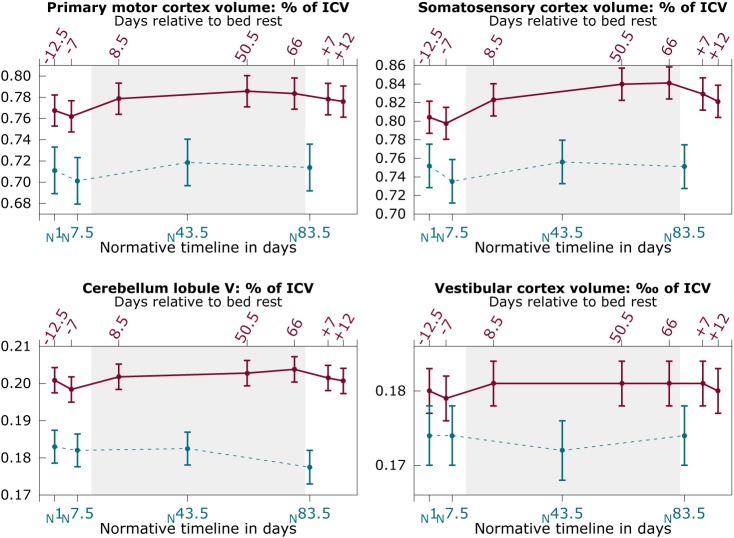
Fig 2. Regional gray matter changes as a function of bed rest.
Solid red lines: HDBR subjects; Dashed blue lines: control subjects; Graphs show mean values over subjects with pooled standard errors. Y-axis shows volume of the region of interest as percentage of the intracranial volume (ICV), except for the vestibular cortex region (4th panel), which shows volume as per mille of the ICV. Top x-axis shows the number of days relative to HDBR; bottom axis shows the time in days for control subjects relative to their first assessment.

