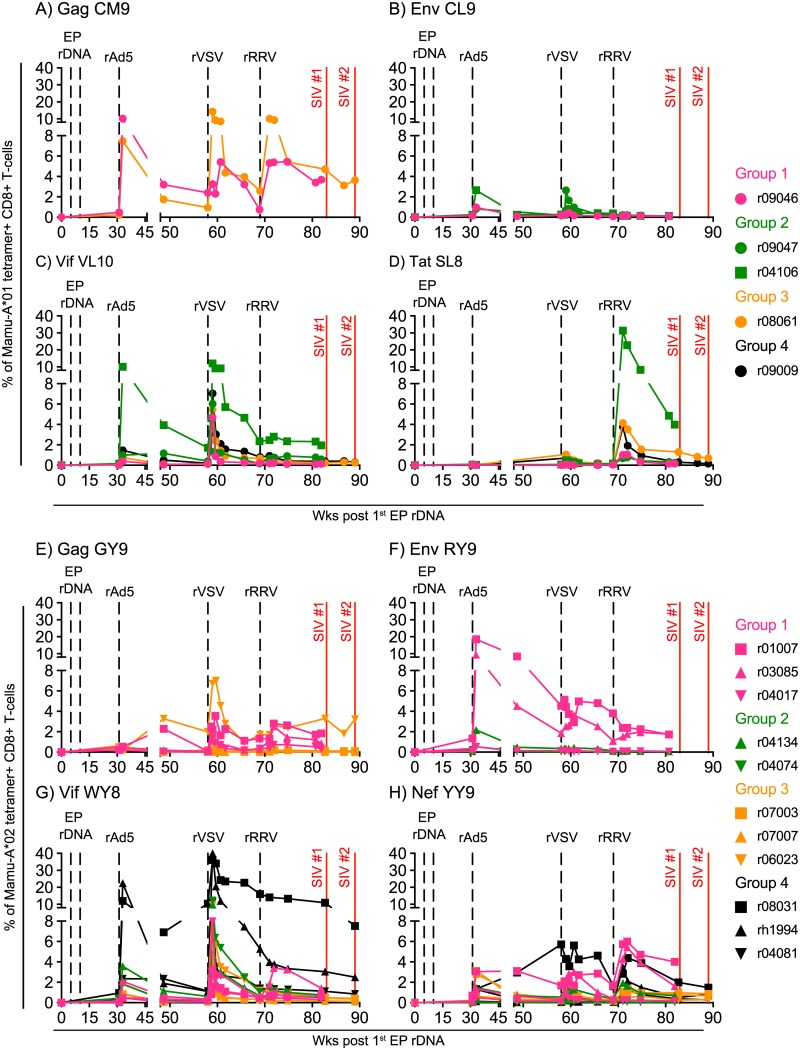
Fig 2. Vaccine-induced CD8+ T-cell responses directed against Mamu-A*01- or Mamu-A*02-restricted epitopes in macaques in Groups 1–4.
Four macaques in each of Groups 1–4 expressed either Mamu-A*01 or Mamu-A*02 (Table 1). Using the appropriate fluorochrome-labeled MHC-I tetramers for each group, we monitored the ontogeny of vaccine-induced CD8+ T-cell responses specific for SIV epitopes restricted by Mamu-A*01 (A-D) and Mamu-A*02 (E-H). Each panel shows the magnitude of vaccine-induced CD8+ T-cells detected by individual MHC-I tetramers in animals in Groups 1–4. The following Mamu-A*01-restricted epitopes were evaluated: A) Gag CM9 (aa 181–189), B) Env CL9 (aa 233–241), C) Vif VL10 (aa 100–109), and D) Tat SL8 (aa 28–35). The following Mamu-A*02-restricted epitopes were evaluated: E) Gag GY9 (aa 71–79), F) Env RY9 (aa 296–304), G) Vif WY8 (aa 97–104), and H) Nef YY9 (aa 159–167). The times of each vaccination (vertical dashed black lines) and when the two SIVmac239 IR challenge rounds were started (vertical solid red lines) are shown in each graph. Macaques in Groups 1, 2, 3, and 4 are color coded in pink, green, beige, and black, respectively.

