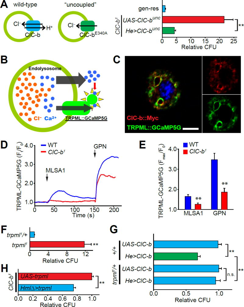
Figure 3. Chloride transporter ClC-b regulates endolysosome luminal Ca2+ levels.
(A) Schematic description of the “uncoupled” mutation and CFU counts from ClC-b1 flies expressing ClC-bunc in macrophages. Values shown are normalized to genomic rescue (gen-res) means. (B) Schematic description of the experiment using TRPML::GCaMP5G to assess endolysosomal Ca2+. (C) Confocal image showing colocalization of the expressed TRPML::GCaMP5G with ClC-b::myc in an isolated larval macrophage. Scale bar represents 5 µm. (D) Representative traces showing changes in TRPML::GCaMP5G fluorescence in larval macrophages isolated from wild-type (blue) and ClC-b1 (red) larvae. Each trace represents mean values from ≥10 macrophages measured in a single experiment. Concentrations of MLSA1 and GPN were 40 µM and 200 µM respectively. (E) Quantification of the endolysosomal Ca2+ measurement experiments. (F) CFU counts from flies of the indicated genotypes. trpml1 flies expressed the trpml+transgene only in neurons to circumvent the pupal lethality observed in trpml-deficient flies. Values shown are normalized to trpml1/+ means. (G) CFU counts from flies overexpressing ClC-b in wild-type trpml1/+ genetic backgrounds. Values shown are normalized to UAS-ClC-b;trpml1/+ means. (H) CFU counts from ClC-b1 flies with or without overexpression of trpml in macrophages. Values are normalized to ClC-b1;UAS-trpml means. All values shown represent mean ±SEM. See also Figure S3.

