Figure 2.

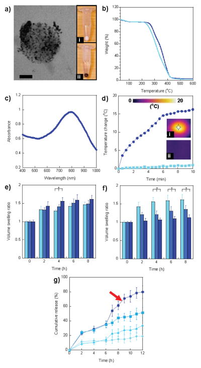
Characterization of multi-targeted nanoparticles. a) TEM image of HPG-Dox-30D70H nanoparticles. Au nanoparticles were homogeneously distributed in nanoparticles. The images of nanoparticle pellets after centrifugation of (i) Dox-30D70H (white) and (ii) HPG-Dox-30D70H (dark brown). Scale bar = 50 nm. b) TGA curves of HPG-Dox-10D90H (light blue) and HPG-Dox-30D70H (dark blue). c) UV-vis spectrum of HPG-Dox-30D70H nanoparticles exhibited a peak at 795 nm. d) Temperature change of HPG-Dox-30D70H in distilled water (5 mg/mL) and nanoparticle-free distilled water after NIR laser irradiation for 10 min. Images were taken using a NIR thermal camera for (i) HPG-Dox-30D70H in distilled water and (ii) only distilled water at 10 min NIR laser irradiation. e) Volume swelling ratios of HPG-Dox-10D90H (light blue), HPG-Dox-20D80H (blue), and HPG-Dox-30D70H (dark blue) in pH 5.5 20mM phosphate buffer at 0, 2, 4, 6, and 8 h. f) Volume swelling ratios of HPG-Dox-30D70H in pH 5.5 (light blue), 6.5 (blue), and 7.4 (dark blue) 20 mM phosphate buffers at 0, 2, 4, 6, and 8 h. g) Cumulative Dox release from HPG-Dox-30D70H (5 mg/mL) in pH 5.5 (square, dark blue curve) and 7.4 (diamond, light blue curve) 20 mM phosphate buffers over 12 h. To evaluate the photothermal effect induced by NIR irradiation, HPG-Dox-30D70H was incubated at pH 5.5 (circle) and 7.4 (triangle) in 20 mM phosphate buffer and then irradiated under a NIR laser for 6 h. The error is the standard deviation from the mean, where n = 3. * is P < 0.05.
