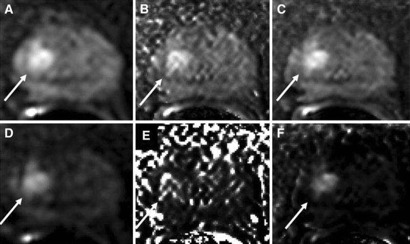
Figure 2.

A 68 year old man with serum PSA of 8.35 ng/mL with apical Gleason 4+3 tumor (10% core involvement). DWI at b=1000s/mm2 (a) acquired at the scanner and calculated using (b) DK model and (c) IVIM model is shown on top row. Similarly DWI at b=2000s/mm2 (d) acquired and calculated using (e) DK model and (f) IVIM model is shown on bottom row.
