
Figure 11.
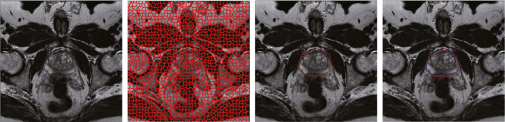
The first image is one original slice of prostate MR. The second image is the corresponding supervoxel map. The third image is the segmentation result of GC. The fourth image is the final segmentation result improved by using AC. The darker contour is the ground truth, while the brighter contour is the final segmentation result. [Color figure can be viewed at wileyonlinelibrary.com]