
Figure 4.
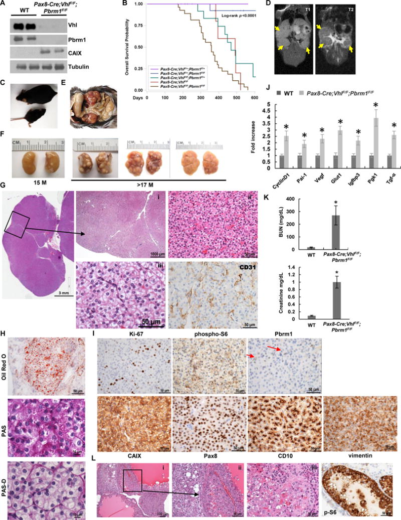
Pax8-Cre;VhlF/F;Pbrm1F/F mice develop ccRCC of lower grade. A, Western blot analysis of the indicated proteins in Pax8-Cre;VhlF/F;Pbrm1F/F kidneys and controls. B, Survival curve of Pax8-Cre;VhlF/F;Pbrm1F/F mice (n=27) compared to Pax8-Cre;VhlF/+;Pbrm1F/+ controls (n=20), and other genotype configurations (Pax8-Cre;VhlF/F;Pbrm1F/+, n=15; Pax8-Cre;Pbrm1F/F, n=5; Pax8-Cre;VhlF/+;Pbrm1F/F, n=17). C, Pax8-Cre;VhlF/F;Pbrm1F/F mice are smaller and runted compared to littermate controls. D, Coronal T1-weighted (left) and T2-weighted (right) images of a Pax8-Cre;VhlF/F;Pbrm1F/F mouse with clearly visible homogeneous tumors with intermediate-to-low signal intensity on T2-weighted images (yellow arrows). E, Kidneys in situ in Pax8-Cre;VhlF/F;Pbrm1F/F mutant mouse. F, Representative macroscopic images of kidneys from Pax8-Cre;VhlF/F;Pbrm1F/F mutant mice at 15 and >17 months of age showing large solid tumors studding the entire cortex. G, Representative H&E microphotographs of kidney sections from Pax8-Cre;VhlF/F;Pbrm1F/F mice showing large ccRCCs completely replacing the kidney. (i) Multiple large solid tumors with pushing borders; (ii) solid tumor with thin arborizing vascular network, monomorphic neoplastic cells with minimal atypia, inconspicuous nucleoli, and mitosis; (iii) prominent cytoplasmic clearing with morphology indistinguishable from low grade human ccRCC. CD31 IHC highlights vasculature. H. Oil Red O staining for triglycerides and neutral lipids, and Periodic acid-Schiff staining without (PAS) and with (PAS-D) diastase highlighting the abundant lipid and moderate glycogen in the neoplastic cells. I, IHC of Ki-67, phospho-S6 (weak and focal), Pbrm1 (lost in tumor nuclei, but retained in normal endothelial cells (red arrows)), CAIX, Pax8, CD10, and vimentin in RCCs of Pax8-Cre;VhlF/F;Pbrm1F/F mice. J, qRT–PCR for the indicated Hif target genes in the kidneys of Pax8-Cre;VhlF/F;Pbrm1F/F mice and controls (n=3 independent mice; PCR done in triplicate). K, Plasma BUN and Creatinine measurements in Pax8-Cre;VhlF/F;Pbrm1F/F mice and controls (n=3 independent mice for each group). L, H&E (i-iii) and phospho-S6 (p-S6) IHC of rare higher grade cystic tumors with pleomorphism and atypia (seen in 4.3% (11/253) of the lesions in 20% of the mice (n=5/26) as well as prominent phospho-S6 resembling those observed in Bap1-targeted kidneys.*, P < 0.05. (WT, age-matched VhlF/F;Pbrm1F/F mice without Cre.)