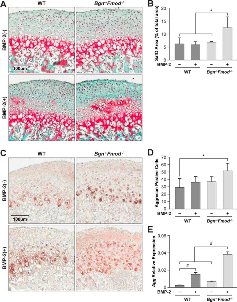
Figure 2.

A) Safranin-O staining of WT (left panels) and Bgn−/−Fmod−/− condyles (right panels) treated without (top panel) or with (bottom panels) BMP-2. B) Histomorphometic quantitation of safranin-O positive area/total area. C) Immunohistochemistry for aggrecan (Acan) in WT (left panels) and Bgn−/−Fmod−/− condyles (right panels) treated without (top panel) or with (bottom panels) BMP-2 D) histomorphic evaluation of Agan positive cells. E) Relative expression of Acan mRNA in WT vs. Bgn−/−Fmod−/− with or without BMP-2. *p<0.05, #p<0.01.
