
Figure 6. MM cells induced-Tregs (iTregs) highly express CD38 and are blocked by Isa.
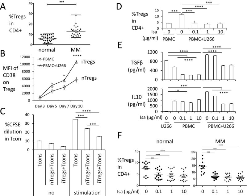
(A) Percentages of Tregs in CD4+ lymphocytes were determined in normal donors (n=27) and MM patients (n=26). Shown are median ± ranges. (B) CD38 levels were examined at indicated time periods in iTregs (triangle) vs. nTreg (square) in PBMC cocultured with or without CD38low/− U266 cells in low dose IL-2-containing culture media. (C) CFSE-labeled Tcons cocultured with either nTregs isolated from normal donors or iTregs isolated from MM cell induction in ex vivo coculture system were stimulated with (stimulation) or without (no) anti-CD3/CD28 beads for 6 d. Proliferation were determined by percentage of CFSE dilution in Tcons. (D) Percentages of iTregs were determined in co-cultures of MM cells with PBMCs (n=4) treated with indicated doses of Isa for 7 days. Shown are Means ± SEMs. (E) Supernatants of ex vivo co-cultures (in D) were assayed for TGFβ (upper panel) and IL10 (lower panel) by ELISA. (F) PBMCs from healthy donors (n=15) and MM patients (n=13) were treated with Isa, and percentage Tregs in CD4+ lymphocytes was measured by flow cytometry analysis. Shown are Means ± SEMs. *P< 0.05, **P< 0.01, ***P< 0.001, ****P< 0.0001