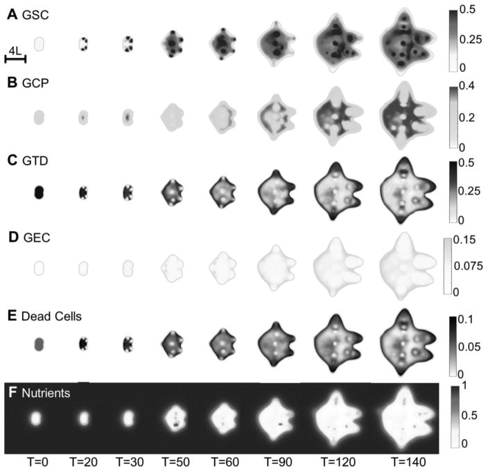
Fig. 4.
2D slices of the untreated vascular tumor in Fig. 2A. (A–H) Distributions of GSCs, GCPs, GTDs, GECs, dead cells, nutrients, vascular-produced GSC promoter (CF) and vessel density at the center of the tumor. Color version in SM. After the vasculature forms, functional vessels release nutrients in the tumor, and several new GSC clusters emerge at the tumor interior. In (D), GEC spontaneously form a network structure in the tumor, as seen in Fig. 2B.

