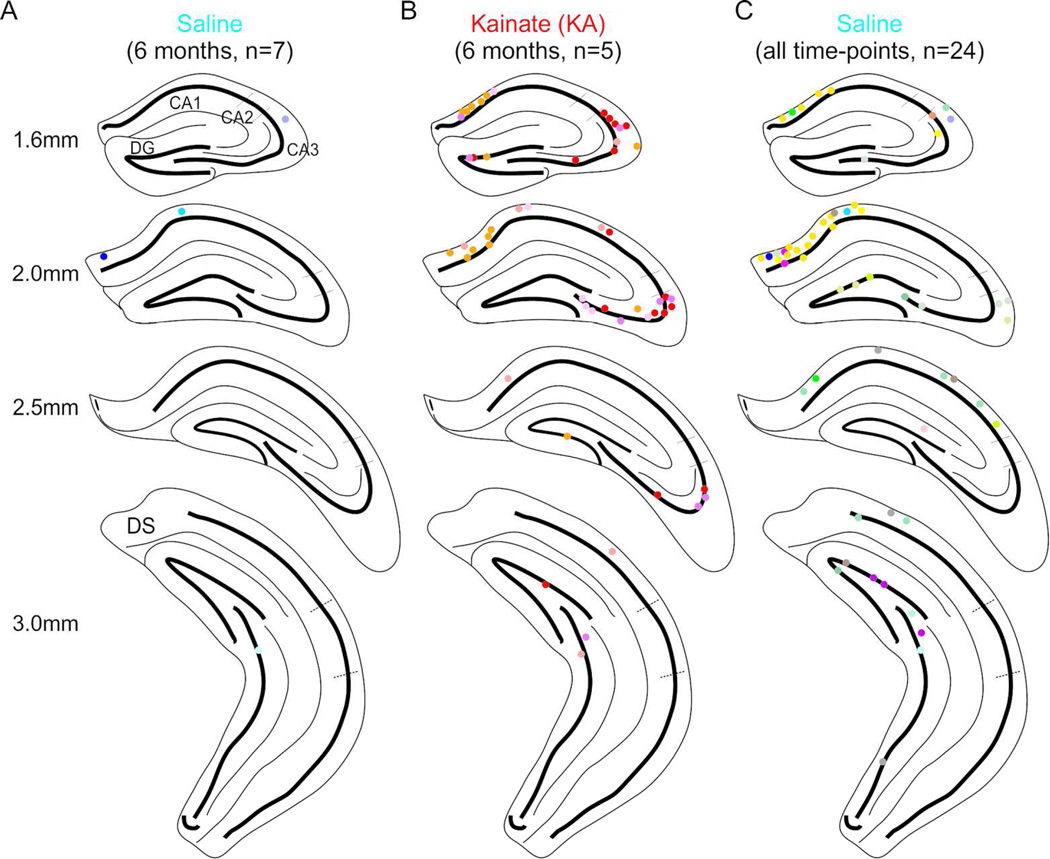
Figure 2. PV+ interneurons co-labeled with FG located throughout the hippocampus.
A–B) PV+ interneurons co-labeled with FG collapsed onto a coronal view of 4 different positions posterior from bregma in saline-injected (A) or KA-injected (B) animals (n = 6 saline, 5 KA) at the 6 month time-point found in CA1, CA3, and the DG. Note the increased number of retrogradely labeled cells following KA (B) compared to saline (A). C) PV+ interneurons co-labeled with FG from all saline-injected animals (n = 24). Each color represents cells from an individual mouse. Dorsal subiculum (DS).

