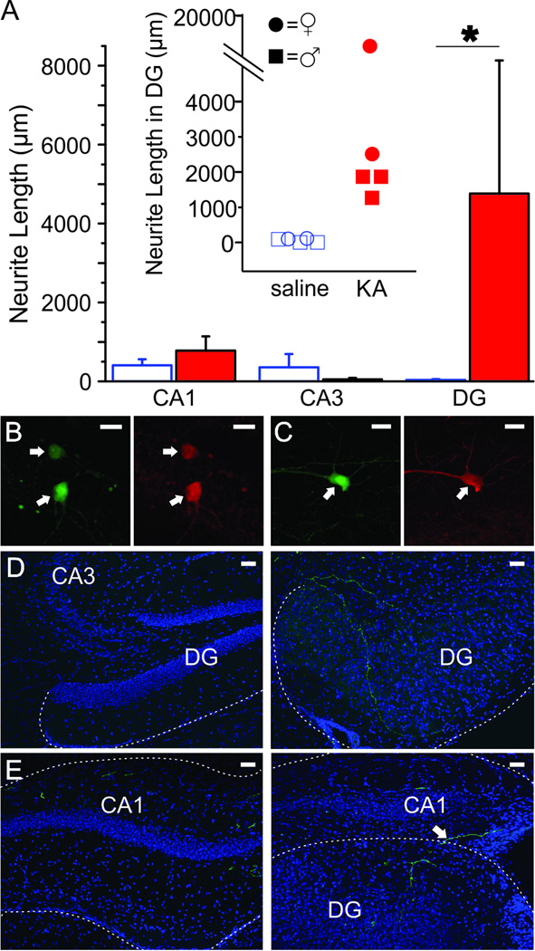
Figure 3. Commissural projections sprout in the DG 6 months following KA injection.
A) Mean length of GFP-labeled fibers located in CA1, CA3, and the DG 6 months post-KA (closed, red bars) or saline (open bars) at 0.84mm left of the sagittal sinus. Asterisk indicates p-value of less than 0.05 (Mann-Whitney, length of neurites located in DG 6 months post-KA vs. 6 months post-saline). Inset shows the length of GFP-labeled fibers in the DG of individual mice 6 months post-KA or saline. Female mice denoted by circles, male mice denoted by squares. There is no apparent sex difference. Overlapping points are offset. B–C) GFP-labeled cells (left panels) in the right (virus-injected) hippocampus are confirmed PV-immunoreactive (right panels) in DG (B) and CA1 (C). Scale bars = 20µm. D–E) In the DG there is significant sprouting of commissural fibers from PV neurons (GFP-labeled fibers) six months after KA-injection (right panels) compared to after saline injection (left panels). Fibers are seen entering the DG from the CA3 region (right panel D) and by crossing the hippocampal fissure (right panel E). Granule cell dispersion is apparent in KA injected animals. Shown with DAPI in blue. Scale bar = 50µm.

