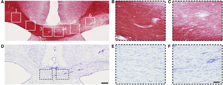
Figure 3.
(A) Representative photomicrograph of myelin-stained sections of the corpus callosum. (B,C) are high magnification images of ROIs 3 and 4 from (A). (D) Representative photomicrographs of Nissl-stained sections of the corpus callosum. (E,F) Are high magnification images of ROIs 3 and 4 in (D). Scale bars: (D,F) 200 and 50 μm, respectively.

