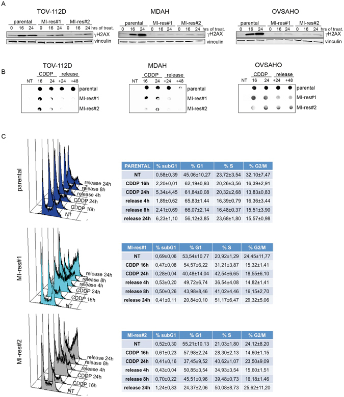
Figure 4.

MI-res cells accumulate less cisplatin-induced DNA damage respect to parental cells. (A) Western blot analyses of S139 phosphorylated Histone H2AX (γH2AX) expression, used as marker of DNA damage, in parental and MI-res cells untreated (0) or treated with cisplatin for 16 and 24 hours, as indicated. Vinculin was used as loading control. (B) Dot blot analyses evaluating the amount of platinated-DNA in parental and MI-res cells untreated (NT) or treated with cisplatin for 16 and 24 hours and then released in cisplatin-free medium for additional 24 (+24) or 48 (+48) hours to allow for DNA repair. (C) FACS analyses of DNA content in TOV-112D parental and MI-res cells untreated (NT) or treated with cisplatin for 16 and 24 hours and then released in cisplatin-free medium for additional 4, 8 or 24 hours, as indicated. A typical histogram for each cell line is shown and the correspondent cell cycle distribution (mean ± SD n = 3 biological replicates) is reported in the lower tables.
