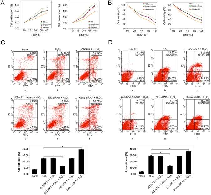
Figure 3.

Anti-apoptotic effect of Kaiso in H2O2 treated endothelial cells. Cells were transfected with pCDNA3.1, pCDNA3.1-Kaiso, NC-siRNA or Kaiso-siRNA and cell viability was analyzed at 0, 12, 24, 36, and 48 h after transfection using CCK8 method (A). Cells were transfected with pCDNA3.1, pCDNA3.1-Kaiso, NC-siRNA or Kaiso-siRNA and were cultured at 37 °C for 48 h. Then, cells were treated with 400 μM H2O2 for 0, 2, 4, 8, and 12 h and cell viability was analyzed at the end of each time period using CCK8 method (B). HUVECs (C) and HMEC-1s (D) were transfected with pCDNA3.1, pCDNA3.1-Kaiso, NC-siRNA or Kaiso-siRNA and were cultured at 37 °C for 48 h. Cells were then treated with 400 μM H2O2 for 8 h and cell apoptosis was examined using Annexin V/Propidium iodide (PI) flowcytometry analysis. The untreated cells served as a blank. Values are presented as mean ± SD, *p < 0.01, compared with H2O2 group, n = 3.
