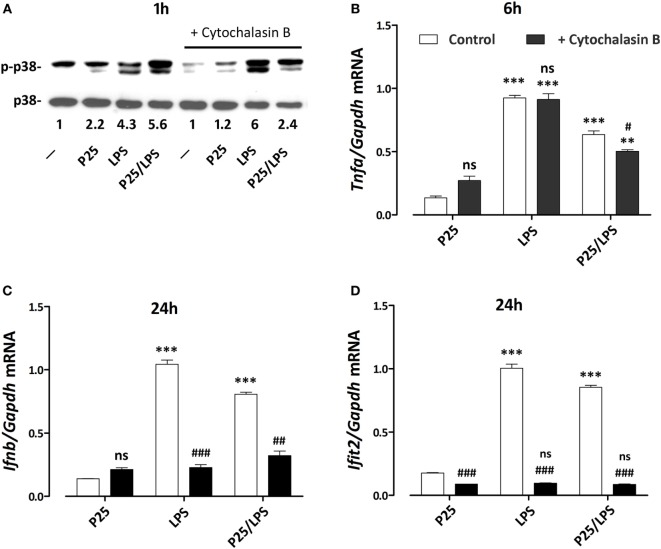
Figure 8.
Effects of cytochalasin B on p38 phosphorylation and pro-inflammatory gene induction. Raw264.7 cells, preincubated for 1 h in the absence or in the presence of cytochalasin B (20 µM), were incubated for the indicated times in the presence of P25 (128 µg/ml), lipopolysaccharide (LPS) (10 ng/ml), or P25/LPS (nominal doses of 128 µg/ml, for TiO2 NP, and 40 pg/ml, for LPS). At the indicated times, the phosphorylation status of p38 (A) and the expression of Tnfa (B), Ifnb (C), and Ifit2 (D) were evaluated by western blot or RT-PCR, respectively. For (A), a representative experiment, performed twice with comparable results, is shown. The numbers represent the quantification of the bands ratio (p-p38/p38) with control cells kept at 1. For (B–D), data are means of two independent experiments, each performed in duplicate, with SD shown. **, ***, p < 0.01 or p < 0.001 vs. corresponding cells (preincubated w/wo cytochalasin B) treated with P25; #, ##, ###, p < 0.05, p < 0.01, p < 0.001 vs. cells under the same experimental condition without cytochalasin B, as evaluated by two-tailed t-test for unpaired data.

