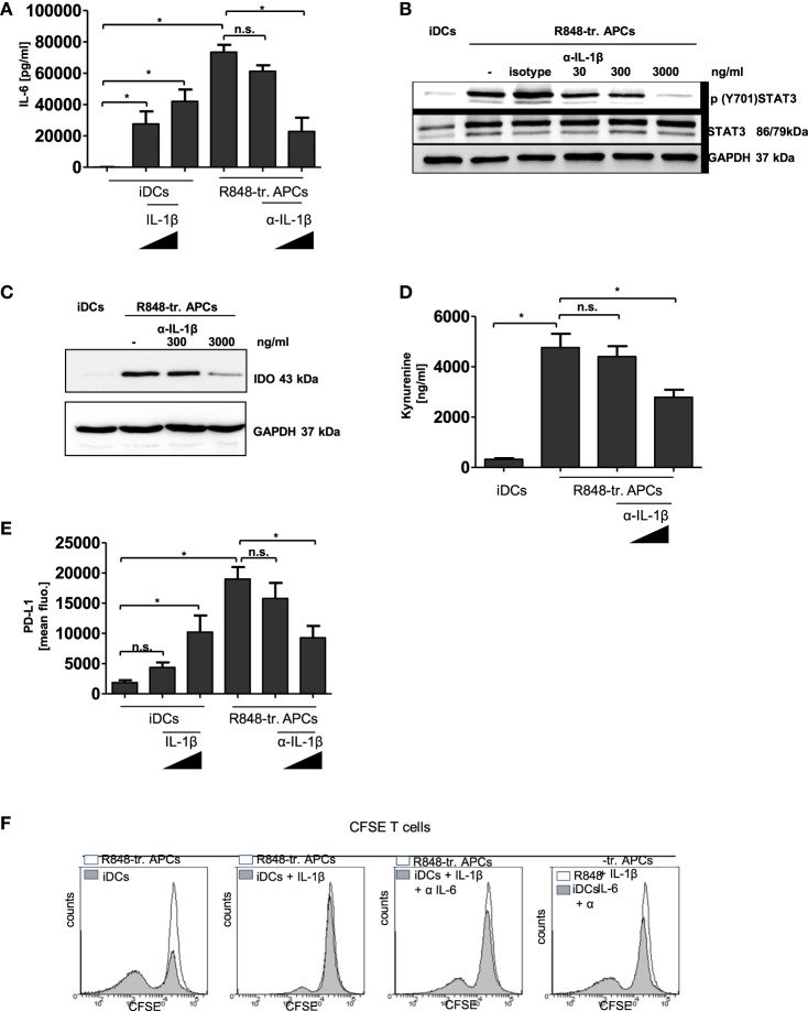
Figure 7.
IL-1β in R848-treated antigen-presenting cell (APC) phenotype. Immature dendritic cells (iDCs) were treated with recombinant IL-1β (100 ng/ml, 1,000 ng/ml). R848-treated APCs were treated with an α-IL-1β neutralizing antibody (300 ng/ml, 3,000 ng/ml) or isotype control (3,000 ng/ml). (A) IL-6 release was measured by enzyme-linked immunosorbent assay (ELISA). (B) Lysates were prepared and adopted in western blot analyses for the detection of p (Y701) signal transducer and activator of transcription 3 (STAT3) and total STAT3. GAPDH served as loading control. (C) Western blot analyses of indoleamine 2,3-dioxygenase. (D) Released Kynurenine was detected in the supernatant of α-IL-1β-treated cells. (D) PD-L1 surface expression was detected by flow cytometry. (A,D,E) show the mean and SD of three donors. ELISAs are performed in duplicates. (F) Monocytes were treated with recombinant IL-1β (3,000 ng/ml) with or without α-IL-6 antibody (1 µg/ml, 10 µg/ml) from the beginning of differentiation into iDCs. After 3 days cells were cocultured with carboxyfluorescein succinimidyl ester (CFSE)-labeled T cells. As control T cells were cocultured with R848-treated APCs. Shown are overlays of flow cytometry data produced with Weasel software.

