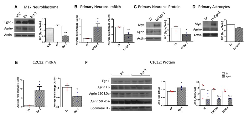
Figure 2.
Human M17 neuroblastoma cells were transiently transfected with pcDNA3.1 and pcDNA3.1Egr-1. (A) Representative western blot from three independent experiments. Egr-1 overexpression led to a significant decrease in agrin protein expression with respect to controls (empty vector, EV). β-actin was used as a loading control. (B) Rat primary hippocampal neurons were infected at 11 days in vitro (11 DIV) with lentiviral control (LV) or lentivirus containing myc-tagged human Egr-1 (LV-Egr-1) at an multiplicity of infection (MOI) of 20. Overexpression of Egr-1 led to a significant increase in EGR1 mRNA expression and decrease in AGRN mRNA expression with respect to LV controls. Values were normalized to GAPDH expression, and results were calculated using the ΔΔCT method and graphed as a fold-change of mRNA expression. n = 3. (C) Overexpression of myc-tagged LV-Egr-1 also led to a significant increase in myc and decrease in agrin protein expression. n = 3. (D) Next, rat primary hippocampal astrocytes were infected at 11 DIV with LV or LV-Egr-1 at an MOI of 20. Egr-1 overexpression did not significantly change agrin protein expression compared to LV controls in these cells. β-actin was used as a loading control. n = 3. (E) Mouse C2C12 myoblast cells were transiently transfected with pCMV6-XL5 (EV) and pCMV6-XL5-Egr-1(Egr-1). Overexpression of Egr-1 led to a significant increase in EGR1 and decrease in AGRN mRNA levels with respect to EV controls. n = 3. (F) When Egr-1 was overexpressed in C2C12 cells, Egr-1 protein levels were increased, and full length, 110 kDa, and 50 kDa agrin protein expression were all significantly decreased compared to EV controls. Coomassie staining was used as a loading control. n = 4. p-values were obtained using Student’s t-tests or 2-way analysis of variance (ANOVA), respectively. *(p ≤ 0.05); **(p ≤ 0.01); ***(p ≤ 0.001); ****(p ≤ 0.0001).

