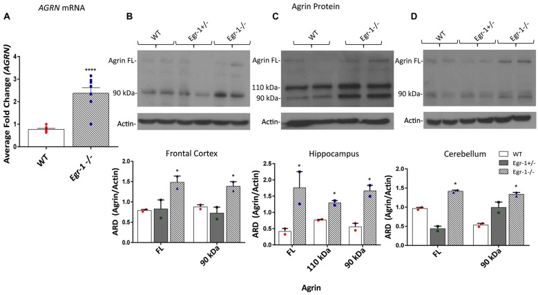
Figure 3.
RNA was extracted from the brains of seven wild type (WT) and seven Egr-1−/− mice, and mRNA was isolated. (A) qPCR showed that Egr-1−/− mouse brain has significantly more AGRN mRNA expression compared with WT mice. Protein samples were prepared from the frontal cortex, hippocampus and cerebellum of six WT, six Egr-1+/− and six Egr-1−/− mouse brains. The western blots presented are representative blots from three independent experiments. (B) Expression of the full length and 90 kDa agrin were significantly increased in the Egr-1−/− frontal cortex compared with that of WT. (C) In the hippocampus, full length, 110 kDa and 90 kDa agrin expression were significantly increased in Egr-1−/− mouse brain compared with WT. (D) In the cerebellum, full length and 90 kDa agrin expression were significantly increased in Egr-1−/− mouse brain compared with WT. p-values were obtained using Student’s t-tests or 2-way ANOVA, respectively. *(p ≤ 0.05); **(p ≤ 0.01); ***(p ≤ 0.001); ****(p ≤ 0.0001).

