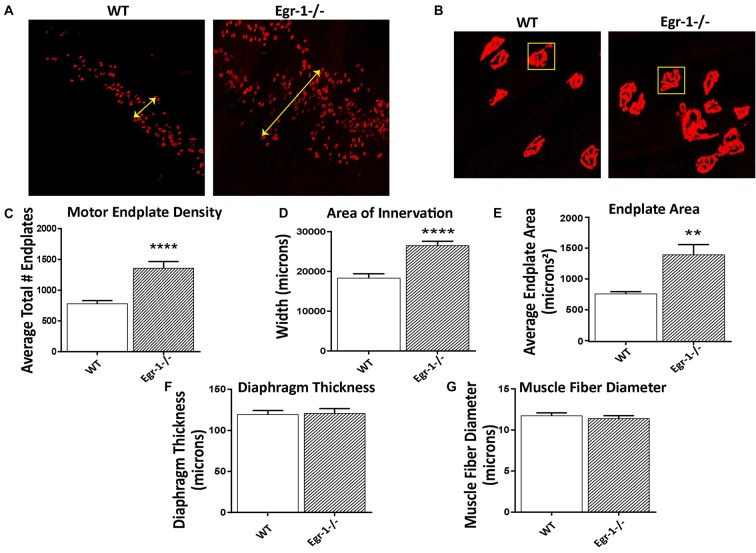
Figure 5.
Diaphragms from five WT and five Egr-1−/− adult mice were whole mounted and stained with α-bungarotoxin to motor endplates made up of AchRs. (A) Representative 10× confocal images of WT and Egr-1−/− mouse diaphragm stained with α-bungarotoxin. (B) Representative 40× confocal images of WT and Egr-1−/− mouse diaphragm stained with α-bungarotoxin. The (C) density (total number) and (D) area of nerve innervation are significantly increased in Egr-1−/− compared with WT. The yellow arrows in the WT and Egr-1−/− panels highlight the width of the innervation band. (E) In Egr-1−/− mouse diaphragm, the area of individual endplates was significantly increased compared to WT. The size difference in the endplates is evident by comparing the identically-sized yellow boxes in the WT and Egr-1−/− panels. There was no significant difference in the thickness of the diaphragm, (F) or in the muscle fiber diameter, (G) in Egr-1−/− mouse diaphragm vs. WT. Z-stack images were quantified and analyzed to obtain results, and analysis was done using ImageJ software. p-values were obtained using Student’s t-tests. *(p ≤ 0.05); **(p ≤ 0.01); ***(p ≤ 0.001); ****(p ≤ 0.0001).

