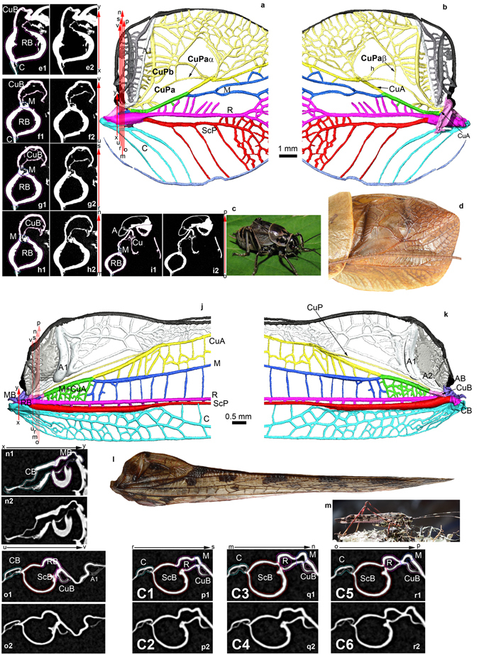
Figure 3.

Wing venation interpretations. (a–i) Prophalangopsidae (Cyphoderris monstrosa). (a) 3D modeling of wing venation using XMT, view from above. (b) 3D modeling of wing venation using XMT, view from below. (c) General habitus (copyright and thanks to D. Gwynne for authorization to use). (d) Complete tegmen (copyright L. Desutter-Grandcolas). (e) Cut xy (1 colored; 2 without color; orientation of tomogram plane XY, tomogram number 1159). (f) Cut uv (1 colored; 2 without color; orientation of tomogram plane XY, tomogram number 1131). (g) Cut rs (1 colored; 2 without color; orientation of tomogram plane XY, tomogram number 1134). (h) Cut mn (1 colored; 2 without color; orientation of tomogram plane XY, tomogram number 1138). (i) Cut op (1 colored; 2 without color; orientation of tomogram plane XY, tomogram number 1112). (j–o) Tettigonioidea (Quiva sp.). (j) 3D modeling of wing venation using XMT, view from above. (k) 3D modeling of wing venation using XMT, view from below. (l) General habitus (copyright and thanks to P.S. Padron for authorization to use). (m) Complete tegmen (copyright L. Desutter-Grandcolas). (n) Cut xy (1 colored; 2 without color; orientation of tomogram plane XZ, tomogram number 130). (o) Cut uv (1 colored; 2 without color; orientation of tomogram plane XZ, tomogram number 207) (p) Cut rs (1 colored; 2 without color; orientation of tomogram plane XZ, tomogram number 223). (q) Cut mn (1 colored; 2 without color; orientation of tomogram plane XZ, tomogram number 232). (r) Cut op (1 colored; 2 without color; orientation of tomogram plane XZ, tomogram number 235). (3D XMT L. Jacquelin).
