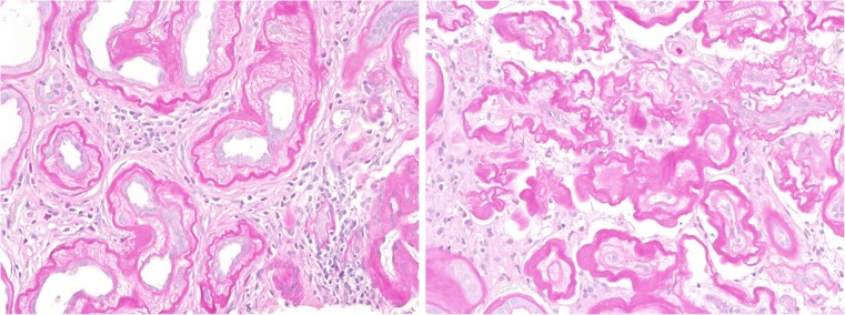
Fig. 1.
Two significantly different types of interstitial fibrosis and tubular atrophy are shown in human renal biopsies. In the left photograph, tubular cells contribute to extracellular matrix increase by a dramatic broadening of the tubular basement membrane. In the right photograph, tubular epithelial atresia with skeleta of tubules is surrounded by interstitial matrix produced by interstitial myofibroblasts. Lesions depicted to the left of Fig. 1 are not seen in mouse, and lesions as seen to the right are seldom observed in mouse (stain: PAS, magnification: 200x)

