Fig. 1.
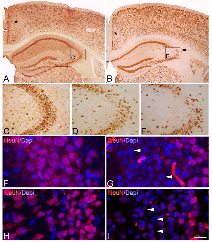
Post-mortem time-related alterations of NeuN immunostaining in mouse cerebral cortex. a, b Low-magnification photomicrographs showing NeuN immunostaining of sections from the neocortex and hippocampus of the brain of mice fixed by immersion immediately after death (PT 0 h, 0 min) (a) or by immersion after 5 h PT (b). Note that in b there are zones with a reduction of NeuN immunostaining in the retrosplenial cortex (asterisk) and the CA3 hippocampal region (arrow). However, in the retrosplenial cortex, these changes were already observed at 0 h (0 min), whereas in CA3, it was observed from 30 min to 5 h. c, d, e High-magnification photomicrographs showing the reduction in the number of NeuN-ir neurons in the stratum pyramidale of CA3 in animals with 30 min PT (d) and, more markedly, with 5 h PT (e) versus immersion-fixed brain tissue that was extracted immediately after death (PT 0 h, 0 min) (c) or perfusion-fixed tissue (see Supplementary Fig. 1S2 A, B). Rectangles in a and b indicate the areas of magnification in c and e, respectively. f–i Confocal images taken from the pyramidal cell layer of CA3 (f, g) and layers II and III of retrosplenial cortex (h, i) of sections immunostained for NeuN and counterstained with DAPI in perfusion-fixed tissue (f, h) and immersion-fixed tissue 5 h PT (g, i). Arrow heads point to DAPI staining of the nucleus of neurons devoid of NeuN immunostaining. Scale bar (in i): 330 µm in (a), B; 80 µm in (c–e); 15 µm in (f–i)

