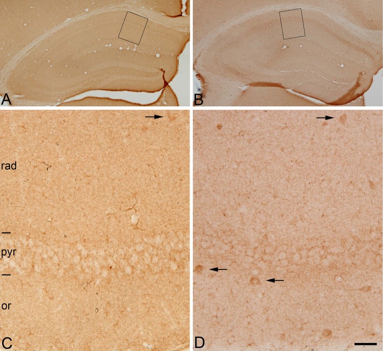
Fig. 3.
Post-mortem time-related alterations of GAD-65-immunostaining in mouse hippocampus. Low (a, b)- and higher (c, d)-magnification photomicrographs (rectangles in a and b, respectively) showing the distribution patterns of GAD-65-immunoreactivity of sections through the hippocampus of mice brains fixed by perfusion (a, c) or by immersion after 5 h PT (b, d). Note that in perfusion-fixed tissue (c) GAD-65 immunoreactivity is present mainly in punctate structures (presumptive axon terminals) distributed in the neuropil or surrounding cell bodies of unlabeled neurons. Occasionally, some cell bodies are also immunostained (arrows). In post-mortem brain tissue, the pattern of immunostaining is similar to that found in perfused brain tissue with the exception that a larger number of GAD-65-ir neurons are stained (d) (arrows). or, stratum oriens; pyr, stratum pyramidale; rad, stratum radiatum. Scale bar: 280 µm in (a, b); 30 µm in (c, d)

